John Condeelis's research interests are in optical physics, cell biology and biophysics, cancer biology and mouse models of cancer. He and his collaborators developed the multiphoton imaging technology and animal models used to identify invasion and intravasation microenvironments in mammary tumors.

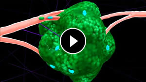
Imaging of live breast tumors at single cell resolution reveals how tumor cells spreadThis led to the discovery of the paracrine interaction between tumor cells and macrophages in vivo, and the role of macrophages in the migration of tumor cells and their dissemination from primary tumors via blood vessels to distant metastatic sites. Based on these results, cell collection techniques, including the in vivo invasion assay, and markers for FACS, were developed for the collection of migrating and disseminating macrophages and tumor cells. This led to the discovery of the mouse and human invasion signatures.
John Condeelis has devised optical microscopes for uncaging, biosensor detection and multiphoton imaging for these studies and has used novel caged-enzymes and biosensors to test, in vivo, the predictions of the invasion signatures regarding the mechanisms of tumor cell dissemination and metastasis. This work has supplied markers for the prediction of breast tumor metastasis in humans. Three of these markers, TMEM, MenaCalc and cofilin x p-cofilin, have been used in retrospective studies of cohorts of breast cancer patients to predict metastatic risk and are now in clinical validation trials. He has authored more than 300 scientific papers on various aspects of cell and cancer biology, biophysics and optical imaging.