Analytical Imaging Facility
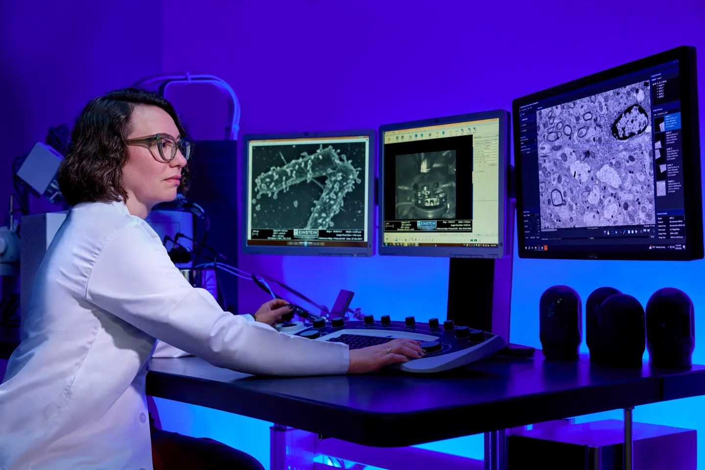
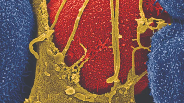
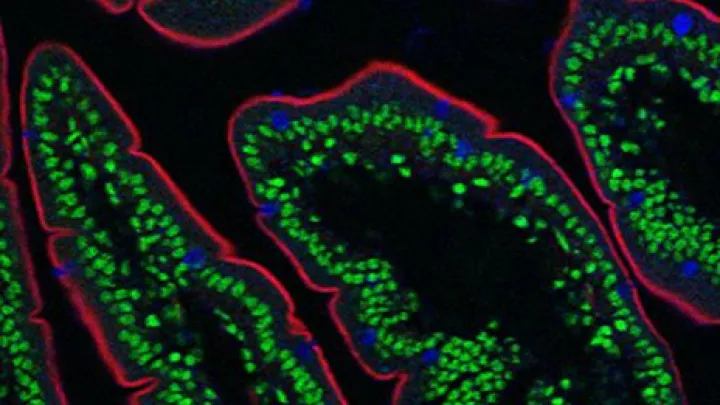
The Analytical Imaging Facility (AIF) is a comprehensive light and electron microscope imaging facility for biomedical scientists with all levels of expertise. The AIF staff is cross trained to seamlessly support visual analysis using techniques ranging from fluorescence light microscope imaging in 3D to high-resolution electron microscopy. This integrated approach facilitates the efficient and appropriate complementary application of these methods for Einstein investigators.
Explore Our Equipment & Services
Contact Us
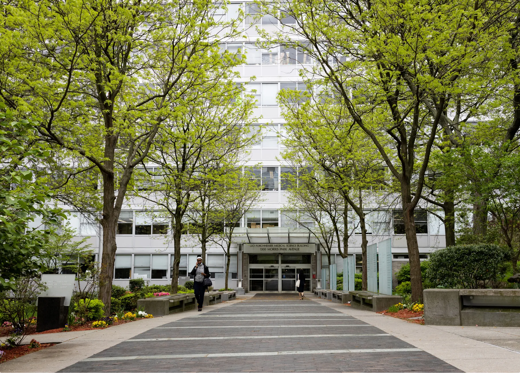
Albert Einstein College of Medicine
1300 Morris Park Ave.
Forchheimer 641
Bronx, NY 10461
718-430-3547
frank.macaluso@einsteinmed.edu
vera.desmarais@einsteinmed.edu
leslie.gunther@einsteinmed.edu
Office Hours
Monday to Friday, 9 a.m.-5 p.m.