

Video could not be played
Research
Research in the CEO lab centers on the clinical problem of metastasis and takes on four areas of focus: 1) Methods for intravital imaging, 2) Understanding the mechanisms of metastasis, 3) Development of biomarkers of metastasis, and 4) Investigating the mechanistic causes of disparities in cancer treatment and outcome.

Video could not be played
Methods for intravital imaging
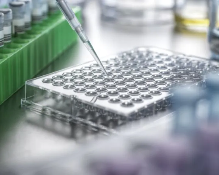
We have pioneered the use of intravital imaging for cancer research. To accomplish this, we have developed 1) Novel microscope instrumentation (how to look), 2) Novel imaging based assays to dissect the live tumor microenvironment (what to look at), and 3) Novel surgical protocols to enable ultra-high-resolution intravital imaging (where to look). This work has required us to develop advanced multiphoton microscopes, and to pioneer the field of Surgical Engineering, where we bring the skills, instruments, and procedures of surgeons into the imaging lab. Techniques we have developed, such as Large-Volume High-resolution Intravital Imaging (LVHR-IVI) allow us to perform long time lapse and serial imaging of a variety of organs, including: mammary glands & tumors, liver, lymphatics and lymph nodes, bone marrow, pancreas, and the lung.
Video could not be played
Understanding the Mechanisms of Metastasis
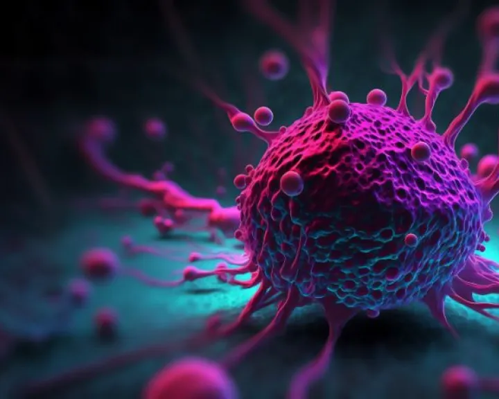
We have used these novel microscopy and intravital imaging techniques to investigate the process of cancer metastasis. Our research has elucidated the mechanisms that underlie how tumor cells metastasize, and revealed the behavior and programming of disseminated tumor cells in secondary sites such as the lymph nodes and the lung. In particular, we have shown that in breast cancer, only a small subset of cancer cells within a tumor become motile and disseminate and that as they do so, these tumor cells pair with macrophages and undergo directed migration toward neo-angiogeneic blood vessels within the bulk of the tumor. Once the tumor cells reach the blood vessels, they cease their movement and combine forces with perivascular macrophages and endothelials to form three-cell portals into the vasculature. These portals, called Tumor Microenvironment of Metastasis (TMEM) doorways, actively open the blood vessels and enable other migratory tumor cells to enter and hematogeneously disseminate. In addition, we have shown that as migratory tumor cells approach TMEM doorways, their interactions with macrophages cause tumor cells to turn on programs of invasiveness, dormancy, and stemness that confer survival and extravasation advantages, as well as chemoresistance. We have termed this programming, the Triple Threat Phenotype (TTP).
Video could not be played
Development of Biomarkers of Metastasis
Since TMEM doorways can be readily identified in preserved (formalin fixed, paraffin embedded) patient tissues, we initiated multiple retrospective clinical trials to determine if the density of TMEM doorways could predict patient outcome. In several independent cohorts, we found that TMEM doorways can predict distant recurrence free survival.
Video could not be played
Investigating the mechanistic causes of disparities in cancer treatment and outcome
Using biophotonics approaches we have discovered the mechanism by which tumor cells are able to gain entry into the blood vascular system and spread to secondary sites.