Case of the Month - April 2023
A 47-year-old man with a recent diagnosis of AIDS and diffuse large B-cell lymphoma was hospitalized because of seizures and an enlarging left neck mass. MRI of the brain showed a 2.5 by 1.2 cm lesion in the left inferior parietal lobe, a 1.4 by 0.7 cm ring-enhancing lesion in the right frontal lobe, and other scattered bilateral lesions. A craniotomy was done, and biopsies obtained from the right temple lesion for aerobic, anaerobic, fungal, and mycobacterial cultures, polymerase chain reaction for Toxoplasma, and surgical pathology.
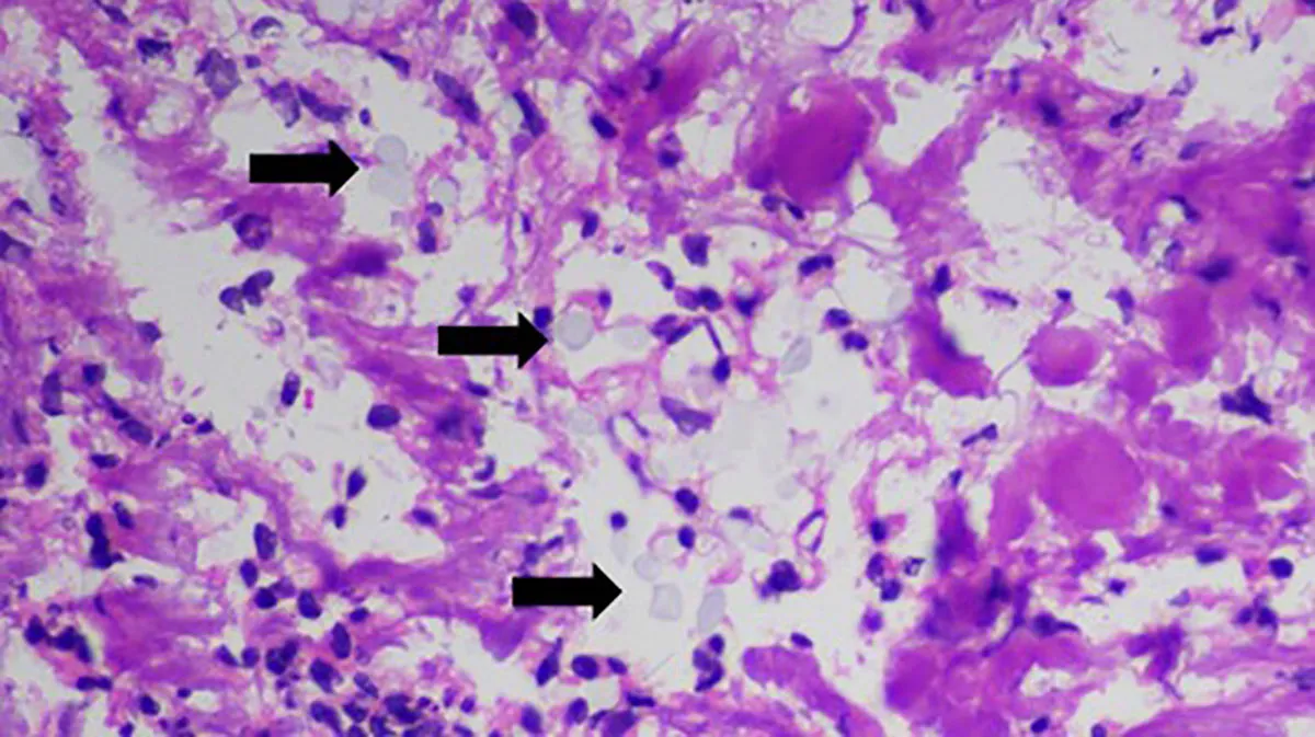
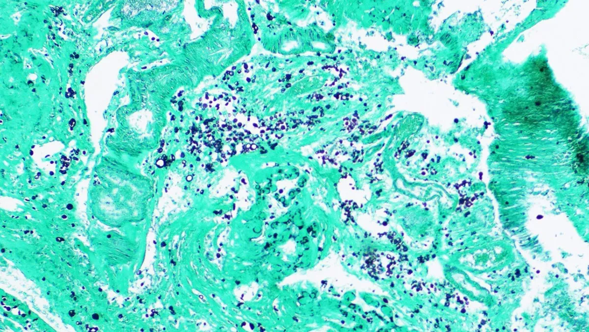
Gram stains, KOH prep, acid-fast stains, and Toxoplasma PCR of the brain tissue were all negative. Representative histopathology slides showing an organism are below.
What is the organism?


