

Gene Imaging
Einstein and Berkeley Receive $4.25 Million from NIH for Next-Generation Gene Imaging
November 2, 2015—(BRONX, NY)—Now that researchers have successfully sequenced the human genome, understanding how those genes operate is one of the next great challenges. To that end, Albert Einstein College of Medicine is taking the lead role in a five-year, $4.25 million grant awarded by the National Institutes of Health (NIH) to develop tools that can image genes and the proteins for which they code.
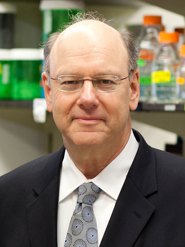
Robert Singer, Ph.D.“Our goal is to develop new instruments to peer into living cells and tissues of animals so that we can visualize cells’ innermost workings in real time—particularly the processes by which their genes are regulated,” said the principal investigator, Robert Singer, Ph.D., professor and co-chair of anatomy & structural biology, co-director of the Gruss Lipper Biophotonics Center and the Integrated Imaging Program, and the Harold and Muriel Block Chair in Anatomy & Structural Biology at Einstein. “These tools include new and more powerful microscopes and fluorescent probes that emit light when imaged. They will reveal the mechanisms that turn genes on and off, which are ultimately at the crux of life itself and the basis of all diseases.” A co-investigator at Einstein, Robert Coleman, Ph.D., is also involved in developing technology for imaging chromatin modifications.
In addition to Einstein, two other institutions are involved in the project: the Janelia Research Campus of the Howard Hughes Medical Institute (HHMI), where Dr. Singer is a senior fellow, and the University of California at Berkeley. HHMI’s Transcription Imaging Consortium is coordinating the efforts of the investigators. The Berkeley component of the grant is led by Xavier Darzacq, Ph.D.
Microscopes that will be specifically adapted for conducting research under the grant include the multifocal microscope, the lattice light sheet microscope, the adaptive optics microscope, and the high-speed three-color super registration microscope, all of which were developed at Janelia.
“Our goal is to develop new instruments to peer into living cells and tissues of animals so that we can visualize cells’ innermost workings in real time—particularly the processes by which their genes are regulated.”
– Robert Singer, Ph.D.
Dr. Singer is a world-renowned expert on mRNA (messenger ribonucleic acid), whose principal role is to carry DNA’s instructions from the cell’s nucleus to the cytoplasm for protein synthesis. His lab has shown that the dynamics of RNA transcription of a single gene can be observed by live-cell imaging and by multiplexed fluorescent probes (probes of different wavelengths). In addition, Dr. Singer has pioneered rapid and sensitive microscopy that can visualize single RNA molecules in living cells in real time and has devised methods to track RNA molecules through their life cycle.
Recent discoveries from Dr. Singer’s lab include pinpointing when and where mRNAs are translated into proteins, using florescent proteins to visualize how memories are formed, and determining the first known mechanism by which cells control the survival of mRNA.
The grant, which was awarded by the National Institute of Biomedical Imaging and Bioengineering, part of the NIH, is titled “Tools for Imaging the Functional Genome in Living Cells and Tissues” (1U01EB021236-01).
Other Top Stories
9/11 World Trade Center Exposure Linked to Heart Disease Among NYC Firefighters
On Becoming a Physician: New Einstein Students Receive White Coats and Stethoscopes
Novel Therapy for Acute Migraine Shows Promise in Phase 3 Clinical Trial
First Complete Wiring Diagram of an Animal's Nervous System
Multimillion Dollar NIH Grant to Help Reduce Opioid Use & Get Care to People Who Need It
NIH Grant Funds $23 Million Study of Diseases Affecting People Living with HIV
New TAILORx Data Guides Adjuvant Therapy in Younger Breast Cancer Patients
Einstein Celebrates Its 61st Commencement
Bolstering Biopsies: Testing Patients' Individual Cells to Guide Treatment
Tablet Blog