FULL STORY
Einstein Researchers Uncover Promising Topical Drug for Diabetes-related Wounds
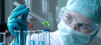
July 28, 2009 — (BRONX, NY) — For those with diabetes, even a small foot wound can heal poorly, leading in some cases to amputation of toes, feet or legs. Now, researchers at Albert Einstein College of Medicine of Yeshiva University and Stanford University have shown that a drug used to remove excess iron from the body reduces by nearly half the time it takes diabetic wounds to heal in mice. This finding could one day help to prevent crippling diabetes complications in humans, including some 71,000 lower-limb amputations that occur each year among Americans with diabetes.
The study is published in the latest online issue of the Proceedings of the National Academy of Sciences (PNAS).
The Einstein and Stanford scientists were led by co-senior authors Michael Brownlee, M.D., professor of medicine and of pathology and the Anita and Jack Saltz Professor of Diabetes Research at Einstein and director of JDRF International Center for Diabetic Complications Research, and Geoffrey Gurtner, M.D., professor of surgery at Stanford University. They based their research on two key findings from previous studies:
|
- The low oxygen level in wounded tissue normally triggers production of vascular endothelial growth factor (VEGF), a protein that stimulates growth of new blood vessels to restore the oxygen needed for healing. But VEGF output and new vessel formation are curtailed in low-oxygen diabetic tissue.
This latest study describes how the scientists were led to the iron-removing drug after first uncovering the molecular basis for poor wound healing in diabetes.
The researchers first assessed how the interaction of low oxygen and elevated glucose affects VEGF levels. They took normal human fibroblasts (cells that secrete fibers to heal wounds) from nondiabetics and grew them in either low-glucose or high-glucose media for four weeks. (The high-glucose condition mimicked the elevated blood-glucose levels of diabetics.) All the cells were then placed in low-oxygen conditions for 24 hours, and VEGF levels were measured.
The fibroblasts cultured in low-glucose conditions increased their VEGF output considerably — by more than 3-fold. By contrast, the high-glucose fibroblasts increased their VEGF production by only 20 percent. These findings were confirmed by experiments using fibroblasts from diabetic patients, and from diabetic and non-diabetic mice: in low-oxygen environments, exposure to high glucose levels was consistently associated with low VEGF production.
The next step was to find the mechanism by which high glucose levels impair VEGF production in diabetic tissues.
The researchers turned their attention to a protein called hypoxia-inducible factor 1a (HIF-1a); under low-oxygen conditions, HIF-1a stimulates VEGF production so that blood flow to oxygen-deprived tissue can be restored. To do its job of turning on VEGF production, HIF-1a needs a partner: a "coactivator" protein known as p300. But in diabetic tissue, the researchers found, HIF-1a and p300 don't always hook up; after culturing fibroblasts under high-glucose conditions, they found that binding between the two proteins was reduced by half.
The problem, Dr. Brownlee and his colleagues discovered, is that high glucose levels inside cells create chemicals called free radicals that release protein-bound iron. This released iron triggers a harmful cascade of chemical reactions leading to dysfunctional p300 proteins that can't effectively bind to HIF-1a.
"We knew that the key thing for improving wound healing in diabetics was to interrupt this chemical cascade," says Dr. Brownlee.
Once they had figured out the molecular pathway responsible for poor wound healing in diabetic tissue, the researchers realized that deferoxamine — an off-patent drug that binds to iron and removes it from the body — might help to short-circuit this pathway. (Deferoxamine has been used for decades to treat diseases such as thalassemia in which toxic levels of iron accumulate in tissues.)
Their experiments with cell cultures suggested that deferoxamine did indeed increase binding between HIF-1a and p300 despite high glucose. Then came the crucial experiments to test whether topical deferoxamine could improve wound healing in diabetic mice. Results were promising; the wounds of diabetic mice treated with the cream healed in 13 days compared with 23 days in untreated diabetic mice.
The next step, says Dr. Brownlee, is to test deferoxamine on human wounds. He notes that when used to remove iron from the body, the drug is given in the form of an injection that can cause side effects ranging from mild stomach upset to rare cases of serious bacterial infections. "But here we're using deferoxamine topically, so the potential for such side effects would be greatly reduced," he notes.
The paper, "The molecular basis for impaired hypoxia-induced VEGF expression in diabetic tissues," appeared in the July 27 online edition of PNAS. In addition to Dr. Brownlee, other Einstein scientists involved in the study were Dachun Yao and Xue-Liang Du.
Other Top Stories
9/11 World Trade Center Exposure Linked to Heart Disease Among NYC Firefighters
On Becoming a Physician: New Einstein Students Receive White Coats and Stethoscopes
Novel Therapy for Acute Migraine Shows Promise in Phase 3 Clinical Trial
First Complete Wiring Diagram of an Animal's Nervous System
Multimillion Dollar NIH Grant to Help Reduce Opioid Use & Get Care to People Who Need It
NIH Grant Funds $23 Million Study of Diseases Affecting People Living with HIV
New TAILORx Data Guides Adjuvant Therapy in Younger Breast Cancer Patients
Einstein Celebrates Its 61st Commencement
Bolstering Biopsies: Testing Patients' Individual Cells to Guide Treatment
Tablet Blog