


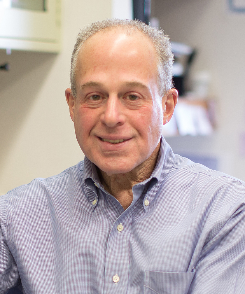
Richard N. Kitsis, M.D.
- Professor, Department of Medicine (Cardiology)
- Professor, Department of Cell Biology
- The Dr. Gerald and Myra Dorros Chair in Cardiovascular Disease
- Director, Wilf Family Cardiovascular Research Institute
Area of research
- Cell Death and Mitochondrial Biology: Fundamental Mechanisms and Roles in Human Disease
Location
- Albert Einstein College of Medicine Jack and Pearl Resnick Campus 1300 Morris Park Avenue Forchheimer Building G46 Bronx, NY 10461
Research Profiles

Professional Interests
Cell Death and Mitochondrial Biology: Fundamental Mechanisms and Roles in Human Disease
The most basic decision that any cell makes is to grow, differentiate, or die. Our laboratory studies fundamental mechanisms of cell death and the roles of cell death in normal biology and human disease. Over a dozen cell death programs have been recognized to date (Cell Death Differ, 2018; Physiol Rev, 2019). Little is understood about how they interconnect mechanistically or functionally. The overarching goal of our work is to create a unified framework of cell death that integrates seemingly “individual” cell death programs. Our hypothesis is that these cell death programs, which often undergo activation in the same cell, are components of an integrated cell death response that has arisen over evolution. We have elucidated basic aspects of cell death biology including mechanisms that mediate apoptosis and regulated forms of necrosis, molecular interconnections among these programs, and connections with other aspects of mitochondrial biology (Mol Cell, 2004; PNAS, 2007; PNAS, 2012; Cell Death Differ, 2014; Nature, 2016; Science, 2018; Nat Commun, 2022; PNAS, 2023; Mol Cell, 2024).
Although our primary focus is basic mechanisms, we are also interested in translational implications. While some of our work has involved models of cancer (Cell Death Differ, 2005; JBC, 2010; Cancer Res, 2011, PLOS One, 2015) and diabetes (Diabetes, 2013; Sci Rep, 2017; Dev Cell, 2021), our primary translational focus is heart disease, the major cause of mortality worldwide. A number of years ago, our lab provided the first evidence that regulated forms of cell death play critical roles in the pathogenesis of myocardial infarction (“heart attack”) (Circulation, 2000; JMCC, 2000; AJP, 2003) and heart failure (JCI, 2003). More recently, the lab has elucidated novel mechanisms of cell death during myocardial infarction (e.g., Circulation (in press)) and developed small molecule drug prototypes as new therapies for heart disease (Nat Cancer, 2020).
Current research in the lab includes:
- Identification of a novel caspase-9-mediated necrosis pathway
Caspase-9 is the sole initiator caspase in the mitochondrial apoptosis pathway. It is activated within the apoptosome, a multiprotein complex in the cytoplasm, which is the only known platform for caspase-9 activation in higher eukaryotes. Caspase-9 then cleaves and activates effector caspases-3 and -7, which hydrolyze hundreds of cellular substrates to bring about apoptotic cell death. We discovered that caspase-9 also mediates necrotic cell death through a distinct mechanism. Caspase-9 enzymatic activity is required for necrosis as it is in apoptosis. However, caspase-9-mediated necrosis is independent of Apaf1, the scaffold of the apoptosome, and independent of caspases-3 and -7, the substrates of caspase-9 that mediate apoptosis. We are currently attempting to identify the platform that activates caspases-9 in necrosis and the downstream pathway. Caspase-9-mediated necrosis appears to operate in multiple cell types. In addition, gene knockout studies show that it is critical for cardiac damage during myocardial infarction.
- Role of necroptosis in myocardial infarction
Necroptosis, a regulated necrosis program defined by RIPK3 → MLKL signaling, is important in myocardial infarction. We have discovered that RIPK3 is activated in cardiomyocytes in this syndrome by ZBP1, a cytoplasmic sensor of double stranded nucleic in the left-handed (or “Z”) conformation - rather than by the RIPK1 as has been widely assumed (Circulation (in press)). This finding provides the initial hint that Z-nucleic acids may play a role in ischemic signaling. We are currently delineating the Z-nucleic acids that bind and activate ZBP1 during myocardial infarction and their subcellular origin (nucleus and/or mitochondria). This may provide an entry point for understanding how ischemia connects with the necroptosis machinery. In addition, we are developing small molecule approaches to inhibit this pathway.
- Regulation of mitochondrial function by cyclophilin D
Cyclophilin D, the sole cyclophilin in mitochondria, is a peptidyl cis-trans prolyl isomerase that resides in the mitochondrial matrix. Cyclophilin D regulates multiple aspects of mitochondrial biology including metabolism and cell death. We have employed two interaction screens (co-immunoprecipitation/mass spectrometry and proximity labelling (TurboID)) to identify substrates of cyclophilin D in mitochondria and are currently delineating how these interactions mediate the effects of cyclophilin D on mitochondrial function.
- Regulation of ferroptosis by OPA1
We have discovered that the inner mitochondrial membrane protein OPA1 is a critical regulator of the sensitivity of cells to undergo ferroptosis, an iron-dependent form of necrotic cell death involving hydroperoxidation of fatty acids in multiple cellular membranes. Unexpectedly, sensitization to ferroptosis by OPA1 is independent of the ability of OPA1 to promote mitochondrial fusion, its canonical function. Rather, OPA1 sensitizes cells to ferroptosis by stimulating the generation of mitochondrial lipid ROS and suppressing an ATF4-mediated integrated stress response that would otherwise inhibit ferroptosis (Molecular Cell, 2024. 84:1-17). We are currently delineating links between ferroptosis and other cell death programs.
- Function of the mitochondrial ATP synthase in vivo
The mitochondrial ATP synthase is responsible for the generation of >90% of ATP in eukaryotic cells. We devised a genetic strategy to deplete this multi-subunit enzyme in heart muscle cells in mice in vivo. We have used this model to study: (a) the necessity of the mitochondrial ATP synthase in maintaining energetics and function in cardiomyocytes, a particularly energy-demanding cell type; and (b) whether the mitochondrial ATP synthase also serves a second function as the mitochondrial permeability transition pore, as has been proposed by others. Regarding this second question, our results indicate that, rather than serving as the mitochondrial permeability transition pore, the mitochondrial ATP synthase is instead a negative regulator of this pore (PNAS, 2023. 120(51):e2303713120).
- ER/SR-mitochondria relationships in regulating necrotic cell death
We have previously created novel peptides and small molecules that conformationally activate or inhibit mitofusins (Nature, 2016. 540(7631):74-79; Science, 2018. 360(6386):336-341), which are large GTPases involved in mitochondrial fusion and ER/SR-mitochondria tethering. We are employing these reagents to perturb the physical distance and overlap between ER/SR and mitochondria to study ER/SR-mitochondria relationships and Ca2+ transfer on cardiomyocyte cell death and infarct size in vivo (Nature Cardiovascular Research (in revision)).
- Interrelationships between cellular senescence and cell death in aging
We have employed a genome-wide CRISPR-based screen and a proteomics screen to identify mediators that determine whether a cell under stress undergoes senescence versus cell death. We are currently characterizing studying these mechanisms in cell culture and in vivo models of aging. We hope to use this information to create new senolytic drugs.