


Video could not be played
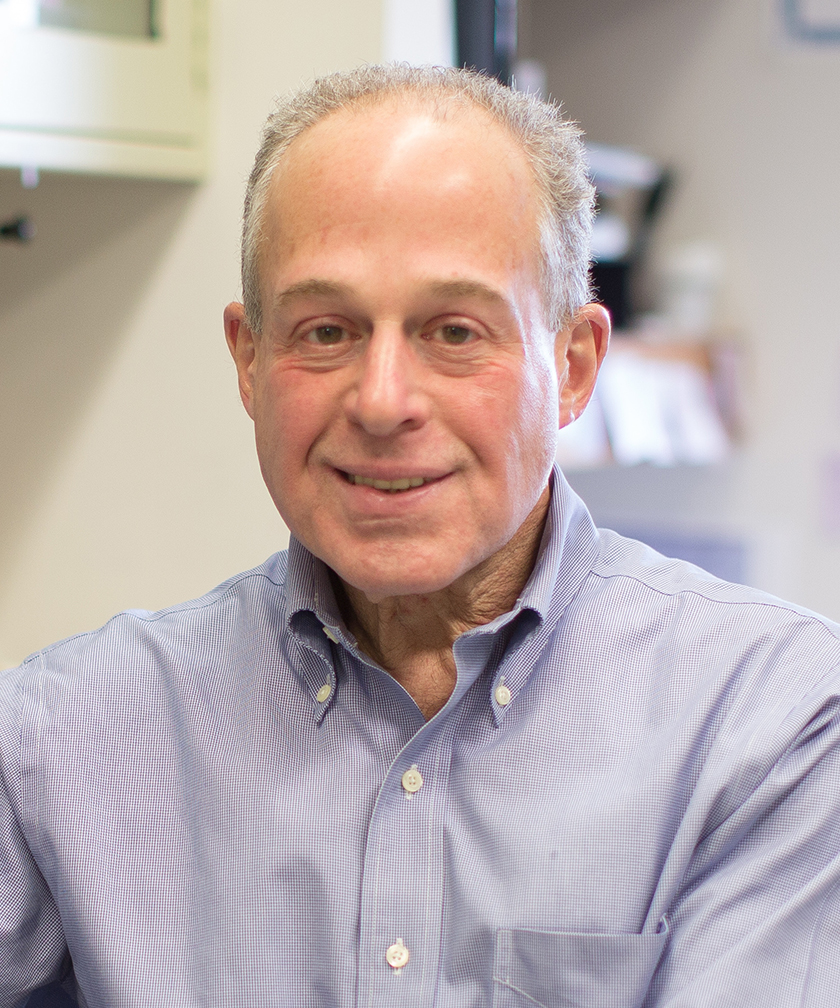
Richard N. Kitsis, M.D.
- Professor, Department of Medicine (Cardiology)
- Professor, Department of Cell Biology
- The Dr. Gerald and Myra Dorros Chair in Cardiovascular Disease
- Director, Wilf Family Cardiovascular Research Institute
Area of research
- Cell Death and Mitochondrial Biology: Fundamental Mechanisms and Roles in Heart Disease
Location
- Albert Einstein College of Medicine Jack and Pearl Resnick Campus 1300 Morris Park Avenue Forchheimer Building G46 Bronx, NY 10461
Research Profiles


Professional Interests
Cell Death and Mitochondrial Biology: Fundamental Mechanisms and Roles in Heart Disease
Past work
The most basic decision that any cell makes is to grow, differentiate, or die. Our laboratory studies fundamental mechanisms of cell death and the roles of cell death in heart disease. I had the opportunity to help establish the field of cardiac cell death research and have focused on this area for the past ~30 years. Although my ultimate goal is to impact cardiac disease, I have taken a basic approach to discovery. Our past work has resulted in the following findings:
Fundamental contributions
- Delineation of a novel class of death-fold interactions (Molecular Cell, 2004)
- Delineation of a signaling pathway through which mitochondrial state is sensed to set cellular sensitivity to ferroptosis (Molecular Cell, 2024)
- Development of the first mitofusin modulators, their structural basis of action, and effects on metabolism and cell death (Nature, 2016; Science, 2018; Nat Commun, 2022).
- Demonstration that the mitochondrial ATP synthase is a negative regulator of the mitochondrial permeability transition pore (PNAS, 2023)
Contributions to understanding cell death in heart and related diseases
- First demonstration that most cell death during reperfused myocardial infarction (MI/R) is mediated by regulated cell death programs (JCI, 1997, Circ Res, 1999; JMCC 2000; Circulation, 2000, 2003, 2003; AJP 2003)
- First demonstration that cardiomyocyte death contributes mechanistically to the pathogenesis of heart failure (JCI, 2003)
- Discovery that BAX, whose canonical function in apoptosis is permeabilization of the outer mitochondrial membrane, promotes Ca2+-induced opening of the inner mitochondrial membrane permeability transition pore, triggering necrosis during MI/R, and the underlying mechanism. (PNAS, 2012)
- Discovery that RIPK3, which mediates necroptosis and pyroptosis during MI/R, is activated by ZBP1 – not by RIPK1 as had been previously assumed – providing the first evidence that Z-conformation (left-handed) nucleic acids are involved in ischemic signaling (Circulation, 2025)
- Discovery of an essential role for BAX in apoptosis, necrosis, and cardiac dysfunction in doxorubicin-induced heart failure and development of the first small molecule allosteric inhibitors of BAX that attenuate doxorubicin-induced cardiomyopathy without compromising anti-cancer efficacy (Nat Chem Biol, 2019; Nat Cancer, 2020)
- Delineation of roles of the death suppressor ARC in heart disease, diabetes, and cancer (PNAS, 2007; JBC, 2007; Circulation 2011; Cancer Res, 2011; Diabetes, 2013; Sci Rep, 2017)
- Delineation of a novel pathway of pancreatic β-cell death in type 2 diabetes (Developmental Cell, 2021)
- Demonstration that peptides which perturb mitofusin 2 conformations can acutely alter ER/SR-mitochondrial spatial relationships in cardiomyocytes to limit infarct size (Nature, 2016; Nat Cardiovasc Res, in revision)
Current Work
Over a dozen cell death programs have been recognized to date. Little is understood about how they interconnect mechanistically or functionally. The overarching goal of our current basic work is to create a unified framework of cell death that integrates seemingly “individual” cell death programs. Our hypothesis is that these cell death programs are components of an integrated cell death response that has arisen over evolution. Our goal is to delineate molecular connections among these cell death programs and to apply this knowledge to understand myocardial infarct generation and develop therapies to reduce infarct size. We are pursuing this theme through the following projects:
1. Mechanism and therapeutic targeting of RIPK3-driven cell death in MI/R
RIPK3 (receptor interacting protein kinase 3) is a direct or indirect activator of the four most important cell death programs that mediate myocardial infarct generation: necroptosis, pyroptosis, ferroptosis, and mitochondrial permeability transition-driven necrosis. As such, RIPK3 is a prime therapeutic target to reduce infarct size, the clinical importance being that infarct size is a major determinant of post-infarct heart failure and sudden cardiac death, the drivers of mortality in ischemic heart disease. Indeed, genetic deletion or pharmacological inhibition of RIPK3 markedly reduces infarct size in mouse models of MI/R. Although RIPK3 may be regulating several relevant cell death programs within the same cardiomyocyte, our single cell analyses suggest the unexpected finding that different cardiomyocytes in the same infarct are killed by activation of different cell death programs. This observation raises the possibility that RIPK3 is activating different cell death programs in different cardiomyocytes. We are currently working to understand this complex cell death signaling because we believe that it will provide critical insights into infarct generation and therapeutic strategies. In addition, we are building upon our finding that ZBP1 (a sensor of Z-conformation (left-handed) nucleic acids), rather than RIPK1 as was previously believed, is the activator of RIPK3 in the context of MI/R (Circulation, 2025). We hypothesize that identification of the specific Z-nucleic acids that bind ZBP1 – enabling ZBP1 to activate RIPK3 – will provide an entry point to dissect the link between ischemia/reperfusion and the cell death machinery. From a therapeutic perspective, we plan to complement our mouse studies showing pharmacological RIPK3 inhibition is cardioprotective using large animal models of MI/R and, pending ongoing safety studies, apply for an IND to conduct first-in-human trials.
2. Identification of a novel CASP9-mediated necrosis pathway
CASP9 (caspase-9) is the sole initiator caspase in the mitochondrial apoptosis pathway. It is activated within the apoptosome, a multiprotein complex in the cytoplasm, which is the only known platform for CASP9 activation in higher eukaryotes. CASP9 then cleaves and activates effector CASP3 and CASP7, which hydrolyze hundreds of cellular substrates to bring about apoptotic cell death. We have discovered that CASP9 also mediates necrotic cell death through a distinct mechanism. CASP9 enzymatic activity is required for necrosis as it is in apoptosis. However, CASP9-mediated necrosis is independent of APAF1, the scaffold of the apoptosome, and independent of CASP3 and CASP7, the substrates of CASP9 that mediate apoptosis. We are currently attempting to identify the platform that activates CASP9 in necrosis, the downstream pathway, and the switch that toggles CASP9 between apoptosis and necrosis. CASP9-mediated necrosis operates in a variety of cell types. Importantly, gene knockout studies show that CASP9 is critical for infarct generation during MI/R, and this involves its necrosis function (independent of APAF1 and CASP3/7). Viable pharmacological strategies exist to specifically inhibit CASP9 activation. We aim to pursue IND-enabling studies to allow testing in humans.
3. Manipulating ER/SR-mitochondria relationships to reduce cardiac damage during MI/R
We have previously developed novel peptides and small molecules that conformationally activate or inhibit mitofusins (Nature, 2016. 540(7631):74-79; Science, 2018. 360(6386):336-341), which are large GTPases involved in mitochondrial fusion and ER/SR-mitochondria tethering. We have employed these reagents to perturb mitofusins and alter the distance and overlap between ER/SR and mitochondria, ER/SR to mitochondria Ca2+ transfer, cardiomyocyte death, and infarct size in the context of MI/R (Nature Cardiovascular Research (in revision)). Using these approaches, we have shown that peptide-mediated mitofusin inhibition reduces infarct size even when administration of the peptide is delayed until reperfusion. We plan IND-enabling studies to allow testing in humans.
4. Molecular determinants of senescence/cell death decisions and implications in aging
Cellular senescence is defined by cell cycle arrest, resistance/altered vulnerabilities to cell death, and an inflammatory/immunogenic secretory program (SASP) in a metabolically active viable cell. Senescence plays important functions in development and wound healing, but excessive senescence load is thought to be a driver of aging through the SASP.We have employed genome-wide CRISPR-based and proteomics screens to identify mediators that adjudicate whether a cell under stress becomes senescent or dies. We have used hits from this screen to identify novel targets for the development of senolytics (drugs that selectively kill senescent cells). In addition, we are studying how various drivers of aging (e.g., lysosomal dysfunction) interact with senescence-cell death decision nodes that we have identified. Finally, most senolytics kill by inducing apoptosis. Recently, we have explored the possibility of utilizing non-apoptotic cell death programs for senolysis. We have found that ferroptosis can be used to selectively kill senescent cells. Conversely, we have discovered that resistance to ferroptosis is important in the acquisition of the senescence state. We are currently delineating the relevant mechanisms.
5. Function of the mitochondrial ATP synthase in vivo
The mitochondrial ATP synthase catalyzes the formation of more than 90% of the ATP in eukaryotic cells. To assess the importance of the mitochondrial ATP synthase for energy generation and cellular function of an energy-demanding cell, we gradually depleted this 28-subunit complex in cardiomyocytes of mice in vivo starting at 5-6 weeks of life. This was accomplished by deleting Atp5l encoding subunit g, one of the supernumerary membrane subunits. Loss of subunit g precludes formation of new mitochondrial ATP synthase complexes. Already existing ATP synthase complexes degrade with a half-life of ~30 days. Mice with cardiomyocyte-specific deletion of Atp5l remain clinically normal for ~12 weeks post-gene deletion, at which time only ~10% of mitochondrial ATP synthase remains. Cardiac systolic function also remains normal, although moderate exercise deficits can be elicited by treadmill testing. Quite unexpectedly, cardiac ATP concentrations also remain normal. We are currently performing radioisotope flux studies to understand the compensatory mechanisms that make this possible. Beginning at 13-14 weeks post-gene deletion, mice begin to demonstrate signs of heart failure, and echocardiography shows marked systolic dysfunction. Strikingly, cardiac ATP concentrations still remain normal. By 16 weeks post-gene deletion, all mice die. We are currently working to elucidate the mechanism of this decompensation, as it does not appear to be mediated by energetic deficits. Our studies thus far indicate activation of a cardiomyocyte death program, which we are currently attempting to causally link with existing metabolic abnormalities. Thus far, the data show that there is tremendous reserve in the mitochondrial ATP synthase and that this complex serves critical functions beyond simply insuring adequate ATP generation. We have also used these mice in other studies to test whether the mitochondrial ATP synthase serves in a second capacity as the mitochondrial permeability transition pore, as proposed by Bernardi and colleagues. We found that, rather than serving as the mitochondrial permeability transition pore, the mitochondrial ATP synthase is instead a negative regulator of this pore (PNAS, 2023. 120(51):e2303713120).