

Video could not be played
Vladislav Verkhusha, Ph.D.
- Professor, Department of Genetics
- Co-Director, Gruss-Lipper Biophotonics Center
Area of research
- Designing synthetic biology and optogenetic tools for manipulation of biological processes across scales, from molecules to mammals. Engineering fluorescent and photochromic probes for all-optical assays, multiplexed microscopy, and deep-tissue imaging.
Phone
Location
- Albert Einstein College of Medicine Jack and Pearl Resnick Campus 1300 Morris Park Avenue Ullmann Building 1217 Bronx, NY 10461
Research Profiles

Professional Interests
My laboratory develops genetically encoded systems for quantitative imaging, sensing, and control of biological processes. We combine optical molecular technologies with target-responsive nanobody-based modules to create programmable platforms that link endogenous molecular activities to engineered cellular outputs. These approaches enable detection, regulation, and computation of intracellular states in living cells and organisms. By integrating molecular engineering, optogenetics, and synthetic biology, we build toolkits for probing and manipulating complex biological systems in physiological and pathological contexts.
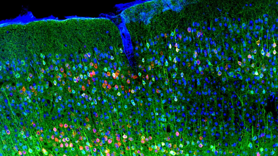
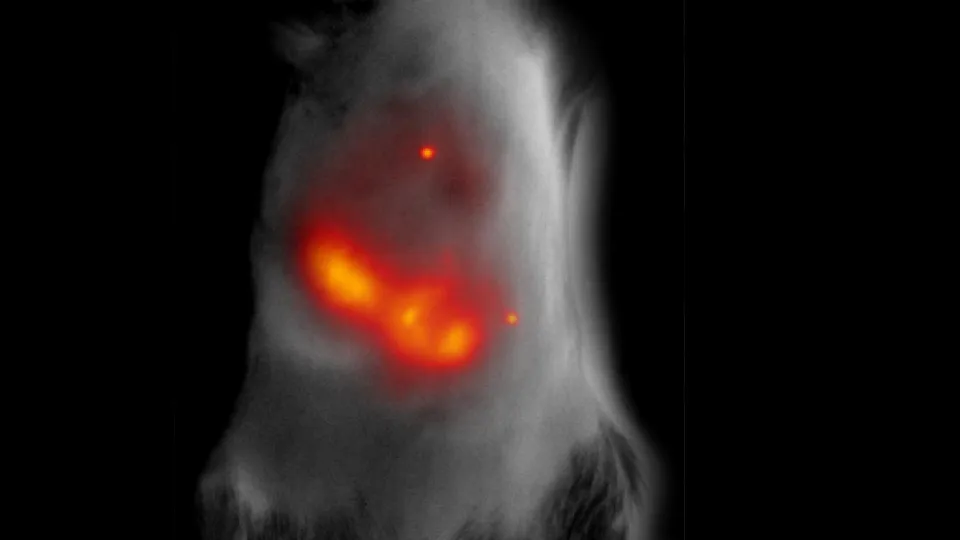
Near-infrared optical probes. We engineer new classes of near-infrared fluorescent probes derived from novel natural photoreceptor templates. Using structure-guided design, directed molecular evolution, and increasingly AI-assisted protein engineering, we generate probes with high brightness, photostability, spectral diversity, and robust performance in mammalian cells and tissues. These probes support multicolor fluorescence microscopy, whole-animal imaging, and integration with hybrid modalities such as photoacoustic imaging.
Biosensors and all-optical assays. Building on photoreceptor scaffolds, we develop functional sensors that report intracellular signaling states, protein-protein interactions, ion dynamics, and enzymatic activity. A particular emphasis is placed on intersectional and logic-based reporters that respond selectively to defined combinations of molecular inputs. Together, these tools enable all-optical assays in which cellular activity can be simultaneously monitored and perturbed, supporting quantitative analysis of complex signaling networks in cells, tissues, and intact organisms.
Optogenetic systems. We design non-opsin optogenetic modules that allow reversible, spatiotemporally precise control of gene expression, protein localization and activity, and intracellular signaling. These systems enable causal interrogation of biological pathways and support experiments that combine optical actuation with near-infrared readouts, extending optogenetics to settings where visible-light tools are limited by scattering or phototoxicity.
Target-stabilizable nanobodies. We develop target-stabilizable nanobody-based platforms that become stabilized only upon binding to specific protein targets or activation states. These systems function as high-contrast intracellular sensors and regulators and serve as programmable input elements for mammalian synthetic biology. By coupling target-dependent stabilization to engineered outputs, they enable the construction of intracellular logic gates and circuits that compute cellular decisions based on target expression or activation profiles.
Programmable systems and application domains. We apply these technologies to engineering and studying complex biological systems, including optogenetic and target-responsive immune cells, oncolytic viruses, and programmable cellular platforms. Additional interests include optogenetic- and target-regulated control of CRISPR/Cas9-based genome editing and transcriptional regulation, regulated production of therapeutic proteins, and approaches relevant to tissue regeneration, neuromodulation, and pain pathways. Collectively, this work provides versatile foundations for translational research in disease-relevant biological systems.
Selected Publications
- Baloban M., Manoilov K.Yu., Karasev M.M., Verkhusha V.V.* and Shcherbakova D.M.* Photoswitchable intein for light control of covalent protein binding and cleavage. Nature Communications 2025, 16:8263. *Co-corresponding authors
- Kasatkina L.A., Ma C., Sheng H., Lowerison M., Menozzi L., Baloban M., Tang Y., Xu Y., Humayun L., Humayun L., Vu T., Song P., Yao J. and Verkhusha V.V. Deep-tissue high-sensitivity multimodal imaging and optogenetic manipulation enabled by biliverdin reductase knockout. Nature Communications 2025, 16:6469.
- Leopold A.V. and Verkhusha V.V. Engineering signalling pathways in mammalian cells. Nature Biomedical Engineering 2024, 8: 1523-1539.
- Barykina N.V., Carey E., Oliinyk O.S., Nimmerjahn A. and Verkhusha V.V. Destabilized near-infrared fluorescent nanobodies enable background-free targeting of GFP-based biosensors for imaging and manipulation. Nature Communications 2024, 15: 7788.
- Oliinyk O.S., Ma C., Pletnev S., Baloban M., Taboada C., Sheng H., Yao. J. and Verkhusha V.V. Deep-tissue SWIR imaging using rationally designed small red-shifted near-infrared fluorescent protein. Nature Methods 2023, 20: 70-74.
- Pennacchietti F., Alvelid J., Morales R.A., Damenti M., Ollech D., Oliinyk O.S., Shcherbakova D.M., Villablanca E.J., Verkhusha V.V. and Testa I. Blue-shift photoconversion of near-infrared fluorescent proteins for labeling and tracking in living cells and organisms. Nature Communications 2023, 14: 8402.
- Leopold AV, Thankachan S, Yang C, Gerashchenko D. and Verkhusha V.V. A general approach for engineering RTKs optically controlled with far-red light. Nature Methods 2022, 19: 871-880.
- Kasatkina L.A., Ma C., Matlashov M.E., Vu T., Li M., Kaberniuk A.A., Yao J. and Verkhusha V.V. Optogenetic manipulation and photoacoustic imaging using a near-infrared transgenic mouse model. Nature Communications 2022, 13: 2813.
- Oliinyk O.S., Baloban M., Clark C.L., Carey E., Pletnev S., Nimmerjahn A. and Verkhusha V.V. Single-domain near-infrared protein provides a scaffold for antigen-dependent fluorescent nanobodies. Nature Methods 2022, 19: 740-750.
- Shemetov A.A., Monakhov M.V., Zhang Q., Canton-Josh J.E., Kumar M., Chen M., Matlashov M.M., Li R., Yang W., Nie L., Shcherbakova D.M., Kozorovitskiy Ye., Yao J., Ji N. and Verkhusha V.V. A near-infrared genetically encoded calcium indicator for in vivo imaging. Nature Biotechnology 2021, 39: 368-377.
- Kaberniuk A.A., Baloban M., Monakhov M.V., Shcherbakova D.M. and Verkhusha V.V. Single-component near-infrared optogenetic systems for gene transcription regulation. Nature Communications 2021, 12: 3859.
- Manoilov K.Y., Verkhusha V.V., and Shcherbakova D.M. A guide to the optogenetic regulation of endogenous molecules. Nature Methods 2021, 18: 1027–1037.
- Redchuk T.A., Karasev M.M., Donnelly S.K., Hülsemann M., Virtanen J., Moore H.M., Vartiainen M.K., Hodgson L. and Verkhusha V.V. Optogenetic regulation of endogenous proteins. Nature Communications 2020, 11: 605.
- Matlashov M.E., Shcherbakova D.M., Alvelid J., Baloban M., Pennacchietti F., Shemetov A.A., Testa I. and Verkhusha V.V. A set of monomeric near-infrared fluorescent proteins for multicolor imaging across scales. Nature Communications 2020, 11: 239.
- Leopold A.V., Chernov K.G., Shemetov A.A. and Verkhusha V.V. Neurotrophin receptor tyrosine kinases regulated with near-infrared light. Nature Communications 2019, 10: 1129.
- Oliinyk O.S., Shemetov A.A., Pletnev S., Shcherbakova D.M. and Verkhusha V.V. Smallest near-infrared fluorescent protein evolved from cyanobacteriochrome as a versatile tag for spectral multiplexing. Nature Communications 2019, 10: 279.
- Shcherbakova D.M., Cammer N.C., Huisman T.M., Verkhusha V.V. and Hodgson L. Direct multiplex imaging and optogenetics of Rho GTPases enabled by near-infrared FRET. Nature Chemical Biology 2018, 14: 591-600.
- Pennacchietti F., Serebrovskaya E.O., Faro A.R., Irina I. Shemyakina I.I., Bozhanova N.G., Kotlobay A.A., Gurskaya N.G., Boden A., Dreier J., Chudakov D.M., Lukyanov K.A., Verkhusha V.V., Mishin A.S., and Testa I. Fast reversibly photoswitching red fluorescent proteins for live-cell RESOLFT nanoscopy. Nature Methods 2018, 15: 601-604.
- Li L., Shemetov A.A., Baloban M., Hu P., Zhu L., Shcherbakova D.M., Zhang R., Shi J., Yao J., Wang L.V. and Verkhusha V.V. Small near-infrared photochromic protein for photoacoustic multi-contrast imaging and detection of protein interactions in vivo. Nature Communications 2018, 9: 2734.
- Redchuk T.A., Kaberniuk A.A. and Verkhusha V.V. Near-infrared light-controlled systems for gene transcription regulation, protein targeting and spectral multiplexing. Nature Protocols 2018, 13: 1121-1136.
- Redchuk T.A., Omelina E.S., Chernov K.G. and Verkhusha V.V. Near-infrared optogenetic pair for protein regulation and spectral multiplexing. Nature Chemical Biology 2017, 13: 633-639.
- Fluegen G., Avivar-Valderas A., Wang Y., Padgen M.R., Williams J.K., Verkhusha V., Cheung J.F., Entenberg D., Castracane J., Keely P., Condeelis J. and Aguirre-Ghiso J. Phenotypic heterogeneity of disseminated tumor cells is preset by primary tumor hypoxic microenvironments. Nature Cell Biology 2017,19: 120-132.
- Yao J., Kaberniuk A.A., Li L., Shcherbakova D.M., Zhang R., Wang L., Li G., Verkhusha V.V.* and Wang L.H.* Multiscale photoacoustic tomography using reversibly switchable bacterial phytochrome as a near-infrared photochromic probe. Nature Methods 2016, 13: 67-73. *Co-corresponding authors
- Shcherbakova D.M., Baloban M., Emelyanov A.V., Brenowitz M., Guo P. and Verkhusha V.V. Bright monomeric near-infrared fluorescent proteins as tags and biosensors for multiscale imaging. Nature Communications 2016, 7: 12405.
- Kaberniuk A.A., Shemetov A.A., and Verkhusha V.V. A bacterial phytochrome-based optogenetic system controllable with near-infrared light. Nature Methods 2016, 13: 591-597.
- Costantini L.M., Baloban M., Markwardt M.L., Rizzo M., Guo F., Verkhusha V.V. and Snapp E.L. A palette of fluorescent proteins optimized for diverse cellular environments. Nature Communications 2015, 6: 7670.
- Shcherbakova D.M. and Verkhusha V.V. Near-infrared fluorescent proteins for multicolor in vivo imaging. Nature Methods 2013, 10: 751-754.
- Piatkevich, K.D., Subach F.V., and Verkhusha V.V. Far-red light photoactivatable near-infrared fluorescent proteins engineered from a bacterial phytochrome. Nature Communications 2013, 4: 2153.
- Subach O.M., Patterson G.H., Ting L.-M., Wang Y., Condeelis J.S. and Verkhusha V.V. A photoswitchable orange-to-far-red fluorescent protein, PSmOrange. Nature Methods 2011, 8: 771-777.
- Koga H., Martinez-Vicente M., Macian F., Verkhusha V.V., and Cuervo A.M. A photoconvertible fluorescent reporter to track chaperone-mediated autophagy. Nature Communications 2011, 2: 386.
- Filonov G.S., Piatkevich K.D., Ting L.-M., Zhang J., Kim K. and Verkhusha V.V. Bright and stable near infra-red fluorescent protein for in vivo imaging. Nature Biotechnology 2011, 29: 757-761.
- Subach F.V., Piatkevich K.D. and Verkhusha V.V. Directed molecular evolution to design advanced red fluorescent proteins. Nature Methods 2011, 8: 1019-1026.
- Bogdanov A., Mishin A., Yampolsky I., Belousov V., Chudakov D., Subach F., Verkhusha V.V. and Lukyanov K.A. Green fluorescent proteins are light-induced electron donors. Nature Chemical Biology 2009, 5: 459-461.
- Subach F.V., Patterson G.H., Manley S., Gillette J.M., Lippincott-Schwartz J. and Verkhusha V.V. Photoactivatable mCherry for high-resolution two-color fluorescence microscopy. Nature Methods 2009, 6: 153-159.
- Gould T.J., Verkhusha V.V. and Hess S.T. Imaging biological structures with fluorescence photoactivation localization microscopy. Nature Protocols 2009, 4: 291-308.
- Subach F.V., Subach O.M., Gundorov I.S., Morozova K.S., Piatkevich K.D., Cuervo A.M. and Verkhusha V.V. Monomeric fluorescent timers that change color from blue to red report on cellular trafficking. Nature Chemical Biology 2009, 5: 118-126.
- Kedrin D., Gligorijevic B., Wyckoff J., Verkhusha V.V., Condeelis J., Segall J.E. and van Rheenen J. Intravital imaging of metastatic behavior through a mammary imaging window. Nature Methods 2008, 5: 1019-1021.
- Gould T.J., Gunawardene M.S., Gudheti M.V., Verkhusha V.V., Yin S.R., Gosse J.A. and Hess S.T. Nanoscale imaging of molecular positions and anisotropies. Nature Methods 2008, 5: 1027-1030.
- Kapoor V., Subach F.V., Kozlov V.G., Grudinin A., Verkhusha V.V.* and Telford W.G.* New lasers for flow cytometry: filling the gaps. Nature Methods 2007, 4: 678-679. *Co-corresponding authors
- Pena P.V., Davrazou F., Shi X., Walter K., Verkhusha V.V., Gozani O., Zhao R. and Kutateladze T.G. Molecular mechanism of histone H3K4Me3 recognition by plant homeodomain of ING2. Nature 2006, 442: 100-103.
- Gurskaya N.G.,# Verkhusha V.V.,# Shcheglov A.S., Staroverov D.B., Chepurnykh T.V., Fradkov A.F., Lukyanov S. and Lukyanov K.A. Engineering of a monomeric green-to-red photoactivatable fluorescent protein induced by blue light. Nature Biotechnology 2006, 24: 461-465. #Co-first authors
- Lukyanov K.A., Chudakov D.M., Lukyanov S. and Verkhusha V.V. Photoactivatable fluorescent proteins. Nature Reviews Molecular Cell Biology 2005, 6: 885-891.
- Chudakov D.M.#, Verkhusha V.V.#, Staroverov D.B., Lukyanov S. and Lukyanov K.A. Photoswitchable fluorescent label for protein tracking. Nature Biotechnology 2004, 22: 1435-1439. #Co-first authors
- Galperin E.#, Verkhusha V.V.# and Sorkin A. Three-chromophore FRET microscopy to analyze multiprotein interactions in living cells. Nature Methods 2004, 1: 209-217. #Co-first authors
- Verkhusha V.V.* and Lukyanov K.A.* The molecular properties and applications of Anthozoa fluorescent proteins and chromoproteins. Nature Biotechnology 2004, 22: 289-296. *Co-corresponding authors