
Video could not be played
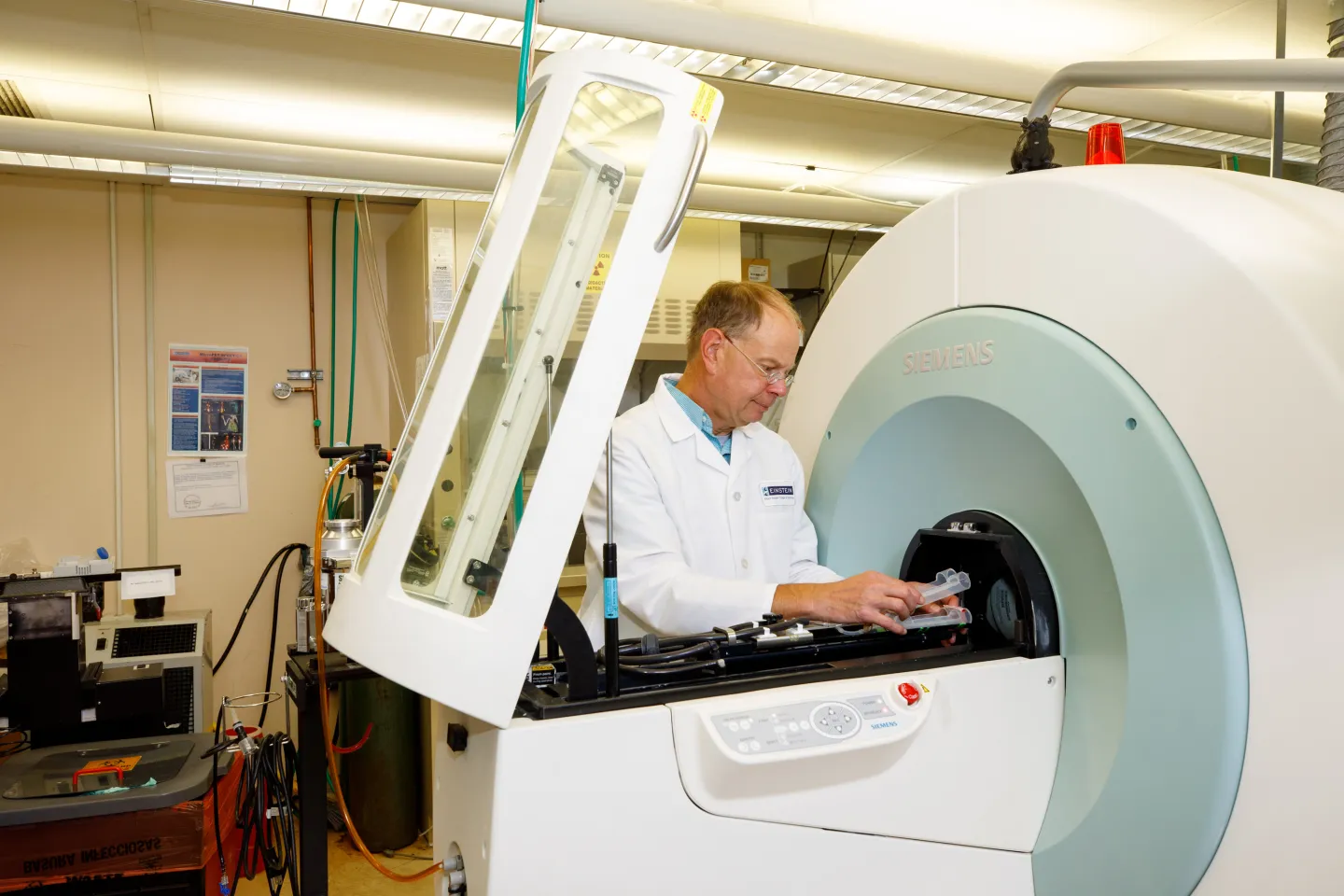
MicroPET Facility
The MicroPET Facility, supported by The M. Donald Blaufox Laboratory for Molecular Imaging and NIH (1S10RR029545 ”MicroPET/SPECT/CT Animal Imaging Device”), and associated with the Gruss MRRC, is designed for pre-clinical investigations using MicroPET (positron emission tomography), SPECT (single photon emission tomography) and CT (computed tomography) examination of small animals to phenotype animal models using a wide variety of radiotracers. Facility staff can train investigators in the use of the tools of MicroPET and help design, implement and interpret all studies. The goal of the facility is to provide investigators with quantitative and high-impact preclinical images.
The following grant must be acknowledged in papers submitted for publication and in abstracts for submission for conferences: NIH S10RR029545 “MicroPET/SPECT/CT Animal Imaging Device" which supported the purchase of the Inveon imaging scanner
Services
- Multiple radiotracers for various phenotyping, for example:
- 18F-Fluorodeoxyglucose (FDG) for analysis of cancers and their response to treatment, cardiac function and infectious disease
- 124I in NIS-expressing tissues
- 64Cu in quantitation of copper metabolism
- Tc99m (MAA) Technetium macro-aggregated albumin for lung perfusion studies used with SPECT imaging
- Tc99m (SC) Technetium sulfur colloid for liver and bone marrow SPECT imaging
- Pilot studies
- Longitudinal studies: scientific/statistical benefit and reduced sacrifice of animals as they serve as their own controls
- Training of investigators in specialized data analysis software to allow independent analysis
Related Shared Facilities
Location and Contacts
MicroPET Facility Gruss 305
David Zhu, Ph.D.
Interim Director, MRRC
Wade Koba, B.S., CNMT
Operations Manager