A Joint Effort Exploring Joint Replacement
With more than a million people receiving artificial hips, knees, elbows, and shoulders each year, joint replacement surgery is one of the world’s most popular orthopedic treatments, bringing greater mobility and pain alleviation to patients with joint problems such as arthritis. But artificial joints can fail over time, and it’s estimated that as many as 15 percent of them eventually do. Thanks to recent research findings of Dr. Laura Santambrogio and Dr. Neil Cobelli, published in Nature Communications, science has a better understanding of why.
Dr. Laura Santambrogio The single most common cause of artificial joint failure is osteolysis. In this process, the constant wear and tear on the weight-bearing surfaces of a joint erodes away tiny particles of debris, such as bits of the polyethylene that makes up portions of a hip prosthesis. These particles provoke an inflammatory response that leads to the breakdown of living bone tissue. Depending on the size of the particle, different types of cells are recruited in an attempt to "digest" the foreign substance, but the ultimate effect is the same. Bone tissue is dissolved, leading to loosening of the implant or even bone breakage.
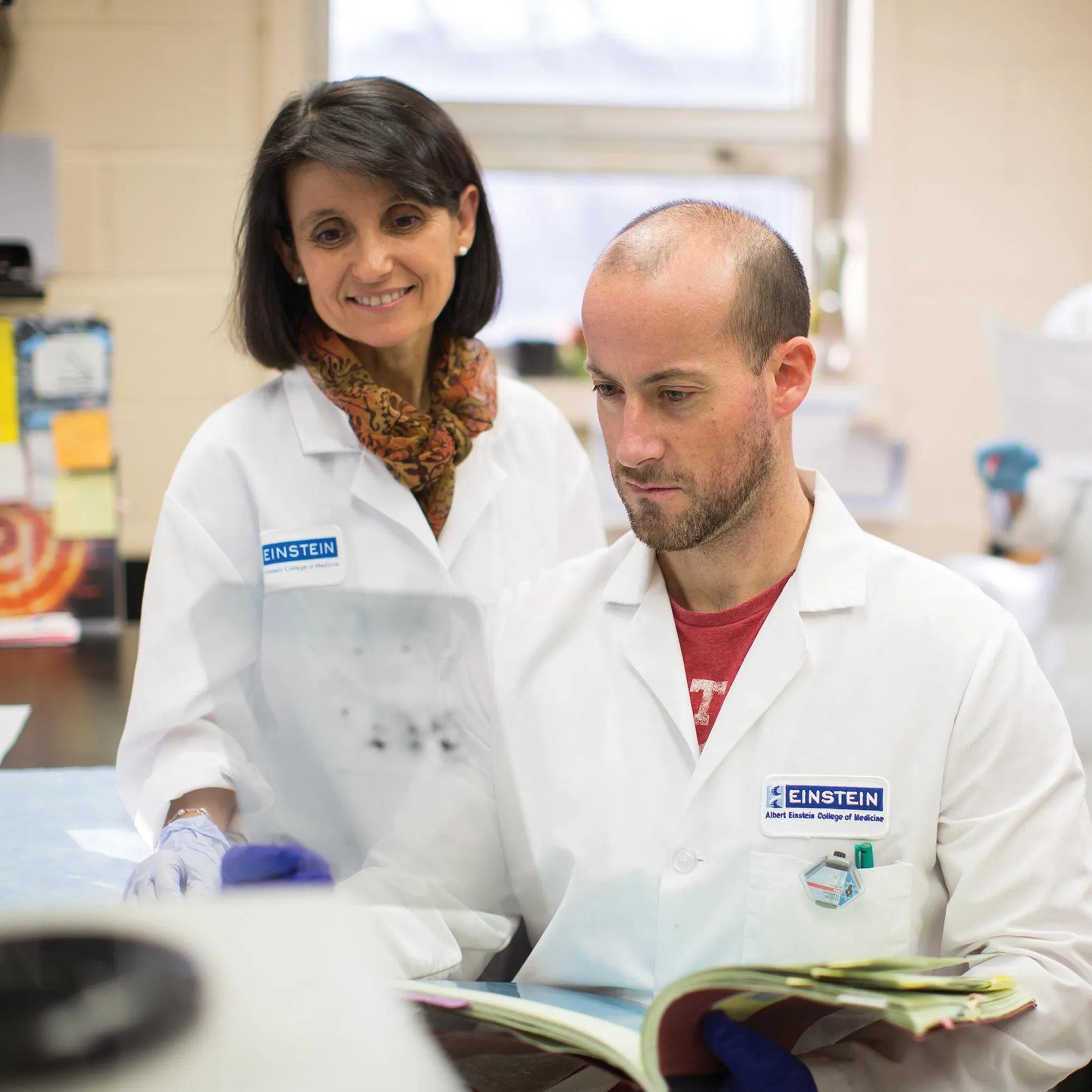
For four years, Dr. Santambrogio, associate professor of pathology and of microbiology & immunology, and Dr. Cobelli, professor of clinical surgery at Einstein and director of orthopedic surgery at Montefiore Medical Center, have worked to describe the multiple cellular pathways of osteolysis. Their work includes an upcoming paper in Nature Communication – with Montefiore director of hematology Dr. Jacob Rand and colleagues – that describes how some damaged cells attempt to repair themselves.
Research like this is important, according to Dr. Santambrogio, because the demand for artificial joints is rapidly growing, and the demands upon the joints themselves are too. "When joint replacement began, patients usually were much older," she said. "Today, replacements are being done when patients are younger and more active."
She added, "Because, 35 to 45 percent of patients undergoing total joint replacement are now under age 65, the implants are under more pressure, and there’s greater likelihood that patients eventually will need new ones."
The first successful hip replacement was performed in 1961. Since then, the substances used in joint replacement have changed in attempts to minimize osteolysis.
"We used to think that the cement used to fix the implant to bone was causing failure, so we eventually started using cement less and less," explained Dr. Cobelli. With or without cement, artificial hips and other joints have included components made of stainless steel, titanium alloys and other metals; ceramics; and plastics such as polyethylene.
So-called "metal-on-metal" joint replacements have been especially controversial, with widespread claims of joint failure and patient hypersensitivity to the metals used. The Food and Drug Administration recently called for an expert review of metal-on-metal hip implants, to help determine whether new safety standards were required. The lesson, according to Dr. Santambrogio, is that no implant material is risk-free. In fact, osteolysis has been associated with every one of them.
Dr. Neil Cobelli "There always will be debris," she said. "But we’re looking for new ideas to reduce them as much as possible. We can modify materials like polyethylene, and there are new types of polymers coming along. But all that begins with understanding what happens at the level of basic science. We need more research on how cells respond to biomaterials."
The very first biomaterials to be implanted, she pointed out, were generally innocuous: dental fillings. But the size and complexity of artificial joints and the invasiveness of joint replacement make it necessary to take a close look at the biocompatibility of the materials used. And that requires both an understanding of the clinical problem and of the most fundamental interactions between the materials and the body.
"This work of Laura, Neil, and others is a great demonstration of how basic science can yield clinically useful information," noted Dr. Michael Prystowsky, professor and chair of pathology, remarking on the collaborative translational research that demonstrates the synergy between Einstein and Montefiore. "In the clinic, at the bedside, you see the problem. But you need basic science to understand it." "This work builds on our combined basic and clinical strengths."
For Dr. Cobelli, the research collaboration also has enhanced his clinical work. "In some ways, I’ve had to learn the language of bench research," he said. "It’s rigorous work. But I feel remarkably rejuvenated as a clinician.
"Putting clinicians and scientists together is beneficial to both, and ultimately to patients as well."
Posted on: Friday, April 20, 2012